
September 22, 2025
The scleral spur
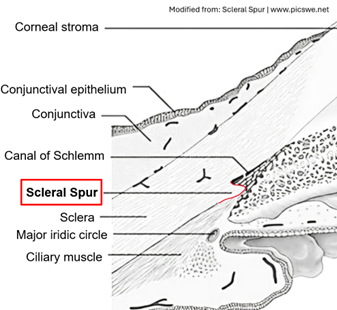
The scleral spur is a ring-shaped structure, primarily collagen with 5% elastin (1), providing structural integrity to the posterior Schlemm's canal. It anchors the trabecular meshwork (anteriorly) and the longitudinal muscle of the ciliary body (posteriorly). Muscle contraction pulls the scleral spur backward, shifting the trabecular meshwork inward. This action widens intertrabecular spaces and expands Schlemm's canal, a continuous dynamic process preventing canal collapse and ensuring proper aqueous humor outflow (2). Clinically, identifying the scleral spur via gonioscopy is essential. It allows differentiation of open-angle from closed-angle glaucoma (3) and precisely targets the anterior trabecular meshwork for procedures like Selective Laser Trabeculoplasty (SLT) and Minimally Invasive Glaucoma Surgery (MIGS).
References
- Moses RA, Grodzki WJ Jr, Starcher BC, Galione MJ. Elastin content of the scleral spur, trabecular mesh, and sclera. Invest Ophthalmol Vis Sci. 1978 Aug;17(8):817-8. PMID: 681140.
- In: Murray A Johnstone. Chapter 3 - Aqueous humor outflow system overview. Editor(s): Robert L Stamper, Marc F Lieberman, Michael V Drake, Becker-Shaffer's Diagnosis and Therapy of the Glaucomas (Eighth Edition), Mosby, 2009, Pages 25-46
- In: https://eyewiki.org/Gonioscopy
Contributor: Francisco Goni