


September 24, 2026
Look for the structural clues of glaucoma
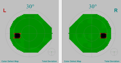
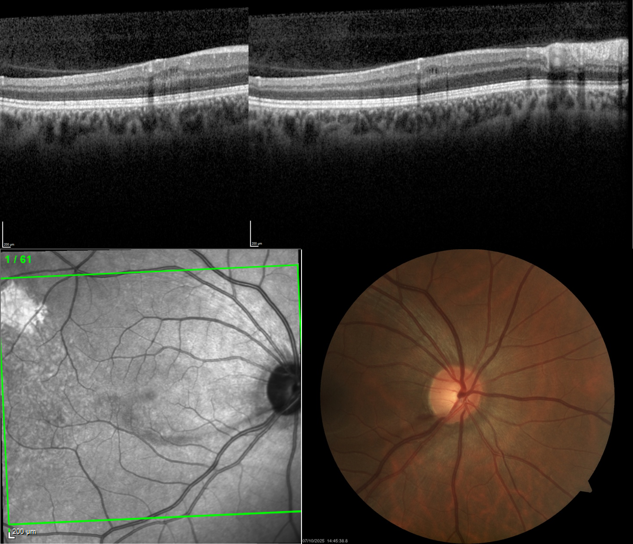
Tip: Glaucoma announces itself through a few recurring structural signs: rim notching, an optic disc (papillary border) haemorrhage, an RNFL defect, a macular ganglion-cell defect and, occasionally, INL microcysts (see imaging examples of a single glaucoma patient below). They need not all be present together, any one may occur in isolation, and a normal visual field does not exclude early glaucoma.
The Science behind the Tip: Glaucoma causes retinal ganglion-cell and axonal loss in characteristic anatomical patterns. Diagnostic confidence increases when abnormalities are topographically concordant across the optic disc, RNFL and macula and, when present, with corresponding visual-field loss. Disc haemorrhages are associated with a higher risk of progression, whereas INL microcysts are uncommon, less specific, and are mainly reported in eyes with more advanced damage.
References
- Pazos M, Traverso CE, Viswanathan A; European Glaucoma Society; Guidelines Task Force; Guidelines Writers, Authors and Contributors; Guidelines Internal Reviewers; Experts by Experience group (patients’ panel); Team of Clinica oculistica of the university of Genoa for medical editing and graphics; External Reviews; EGS Executive Committee; Board of The EGS Foundation. European Glaucoma Society - Terminology and guidelines for glaucoma, 6th Edition. Br J Ophthalmol. 2025 Sep 8;109(Suppl 1):1-212. doi: 10.1136/bjophthalmol-2025-egsguidelines. PMID: 41026937.
- Hasegawa T, Akagi T, Yoshikawa M, Suda K, Yamada H, Kimura Y, Nakanishi H, Miyake M, Unoki N, Ikeda HO, Yoshimura N. Microcystic Inner Nuclear Layer Changes and Retinal Nerve Fiber Layer Defects in Eyes with Glaucoma. PLoS One. 2015 Jun 12;10(6):e0130175. doi: 10.1371/journal.pone.0130175. PMID: 26066021; PMCID: PMC4467090.
- Jung KI, Ryu HK, Oh SE, Shin HJ, Park CK. Thicker Inner Nuclear Layer as a Predictor of Glaucoma Progression and the Impact of Intraocular Pressure Fluctuation. Journal of Clinical Medicine. 2024; 13(8):2312. https://doi.org/10.3390/jcm13082312
Contributor: Jan Van Eijgen, MD PhD, FEBO
Research Group of Ophthalmology (RGO), Department of Neurosciences, KU Leuven, Leuven, Belgium
Department of Ophthalmology, University Hospitals UZ Leuven, Leuven, Belgium

Tip of the Month Manager: Karin Øyo-Szerenyi
Tip of the Month Editors: Karl Mercieca, Francisco Goñi
Tip of the Month Editors in Chief: Manuele Michelessi, Francesco Oddone